Details

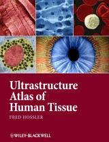
Ultrastructure Atlas of Human Tissues
1. Aufl.
|
230,99 € |
|
| Verlag: | Wiley-Blackwell |
| Format: | |
| Veröffentl.: | 02.04.2014 |
| ISBN/EAN: | 9781118812105 |
| Sprache: | englisch |
| Anzahl Seiten: | 968 |
DRM-geschütztes eBook, Sie benötigen z.B. Adobe Digital Editions und eine Adobe ID zum Lesen.
Beschreibungen
<p><i>Ultrastructure Atlas of Human Tissues</i> presents a variety of scanning and transmission electron microscope images of the major systems of the human body. Photography with the electron microscope records views of the intricate substructures and microdesigns of objects and tissues, and reveals details within them inaccessible to the naked eye or light microscope. Many of these views have significance in understanding normal structure and function, as well as disease processes. This book offers a unique and comprehensive look at the structure and function of tissues at the subcellular and molecular level, an important perspective in understanding and combating diseases.</p> <p>• Presents the major systems of the human body through scanning and transmission electron microscope images</p> <p>• Has images prepared almost exclusively from human tissues</p> <p>• Includes electron micrographs of common pathologies such as fibrotic and emphysemic lung, kidney stones, sickle cell anemia, and skin parasites</p> <p>• Contains sets of 3D images in most chapters</p>
<p>Preface xi</p> <p>Acknowledgments xiii</p> <p>Note to readers xiv</p> <p><b>I. Cellular organelles and surface specializations 1</b></p> <p>A. Nuclei and nucleoli 1</p> <p>B. Mitochondria 6</p> <p>C. Golgi complex 8</p> <p>D. Rough endoplasmic reticulum and smooth Endoplasmic reticulum 10</p> <p>E. Lysosomes 12</p> <p>F. Cytoplasmic inclusions 13</p> <p>G. Plasma membrane junctions 16</p> <p>H. Microvilli 18</p> <p>I. Cilia and centrioles 21</p> <p>J. Plasma membrane infoldings 25</p> <p><b>II. Blood cells 27</b></p> <p>A. Blood composition 27</p> <p>B. Red blood cells 39</p> <p>C. Sickle cell anemia 39</p> <p>D. Granular leukocytes—Neutrophils, Eosinophils, and Basophils 44</p> <p>E. Nongranular leukocytes—Lymphocytes and Monocytes 55</p> <p>F. Blood platelets—Blood clots 70</p> <p><b>III. Connective tissues 77</b></p> <p>A. Composition 77</p> <p>B. Resident cells—Fibroblasts, Adipocytes, Mast cells 90</p> <p>C. Blood cell derivatives—Neutrophils, Eosinophils, Lymphocytes, Macrophages, and Plasma cells 125</p> <p>D. Loose connective tissue example: Lamina propria 132</p> <p>E. Dense irregular connective tissue examples: Dermis and Capsules of organs 132</p> <p>F. Dense regular connective tissue 132</p> <p>G. Cartilage—Hyaline cartilage and Fibrocartilage 144</p> <p>H. Bone—Compact bone and Cancellous bone 161</p> <p><b>IV. Muscle tissues 179</b></p> <p>A. Overview 179</p> <p>B. Smooth muscle—Wall of colon, Branched fibers in wall of ureter, Walls of blood vessels 179</p> <p>C. Skeletal muscle 201</p> <p>D. Cardiac muscle—Atrium 227</p> <p><b>V. Nerve tissues 253</b></p> <p>A. Overview 253</p> <p>B. Peripheral nerves—Optic nerve, Sciatic nerve, Nerve in wall of colon, Myelinated and Unmyelinated nerves 253</p> <p>C. Central nervous system—Cerebrum 287</p> <p><b>VI. Cardiovascular system 303</b></p> <p>A. Overview 303</p> <p>B. Arteries 303</p> <p>C. Veins 328</p> <p>D. Capillaries 336</p> <p>E. Heart-atrium 354</p> <p><b>VII. Lymphatic tissues 359</b></p> <p>A. Overview 359</p> <p>B. Spleen 361</p> <p>C. Thymus 365</p> <p>D. Lymph nodes, lymph nodules/diffuse lymphatic tissue 365</p> <p>E. Tonsils 374</p> <p><b>VIII. Gastrointestinal tract 385</b></p> <p>A. Oral cavity 385</p> <p>B. Overview of the alimentary canal 405</p> <p>C. Esophagus 405</p> <p>D. Stomach 408</p> <p>E. Small intestines—Duodenum, Jejunum, and Ileum 441</p> <p>F. Large intestine (colon) and appendix 474</p> <p><b>IX. Liver and gall bladder 507</b></p> <p>A. Liver 507</p> <p>B. Gall bladder 520</p> <p><b>X. Pancreas 543</b></p> <p><b>XI. Respiratory tract 561</b></p> <p>A. Overview 561</p> <p>B. Trachea, bronchi, and bronchioles 562</p> <p>C. Lungs 579</p> <p><b>XII. Urinary tract 609</b></p> <p>A. Overview 609</p> <p>B. Kidney 609</p> <p>C. Ureters, bladder, and urethra 656</p> <p><b>XIII. Skin 671</b></p> <p>A. Overview 671</p> <p>B. Epidermis 671</p> <p>C. Dermis and hypodermis 725</p> <p>D. Skin parasites 743</p> <p><b>XIV. Eye 749</b></p> <p><b>XV. Ear 797</b></p> <p>A. Overview 797</p> <p>B. Middle ear 797</p> <p>C. Inner ear 802</p> <p><b>XVI. Male reproductive system 819</b></p> <p>A. Testis and epididymis 819</p> <p>B. Vas deferens 841</p> <p>C. Seminal vesicle 844</p> <p>D. Prostate gland 849</p> <p>E. Bulbourethral glands, Glands of littre, and The penis 856</p> <p><b>XVII. Female reproductive system 857</b></p> <p>A. Overview and ovary 857</p> <p>B. Oviduct 860</p> <p>C. Uterus and cervix 873</p> <p>D. Vagina 888</p> <p>E. Placenta 896</p> <p>F. Mammary gland (inactive) 903</p> <p><b>XVIII. Thyroid, parathyroid, and adrenal Glands—examples of endocrine organs 911</b></p> <p>A. Thyroid gland 911</p> <p>B. Parathyroid glands 924</p> <p>C. Adrenal glands 930</p> <p>References 941</p> <p>Index 943</p>
<b>Fred E. Hossler, PhD</b>, is Professor Emeritus of Biomedical Sciences at J.H. Quillen College of Medicine, East Tennessee State University, USA.
<p><i>Ultrastructure Atlas of Human Tissues</i> presents a variety of scanning and transmission electron microscope images of the major systems of the human body. Photography with the electron microscope records views of the intricate substructures and microdesigns of objects and tissues, and reveals details within them inaccessible to the naked eye or light microscope. Many of these views have significance in understanding normal structure and function, as well as disease processes. This book offers a unique and comprehensive look at the structure and function of tissues at the subcellular and molecular level, an important perspective in understanding and combating diseases.</p> <p>• Presents the major systems of the human body through scanning and transmission electron microscope images</p> <p>• Has images prepared almost exclusively from human tissues</p> <p>• Includes electron micrographs of common pathologies such as fibrotic and emphysemic lung, kidney stones, sickle cell anemia, and skin parasites</p> <p>• Contains sets of 3D images in most chapters</p>
Diese Produkte könnten Sie auch interessieren:

The Neural Crest and Neural Crest Cells in Vertebrate Development and Evolution

von: Brian K. Hall

149,79 €


